 “A comparative study of peripheral nerve MRI and ultrasound in MMN and ALS” has been published in Muscle & Nerve. This research was supported in part by JPND through the STRENGTH project, selected for support in the 2012 risk factors call, and the SOPHIA project, selected for support in the 2011 biomarkers call.
Tag Archives: Biomarkers
 “Cerebrospinal α-synuclein in α-synuclein aggregation disorders: tau/α-synuclein ratio as potential biomarker for dementia with Lewy bodies” has been published in the Journal of Neurology. This research was supported in part by JPND through the DEMTEST project, selected in the 2011 biomarkers call.
 “Measurement of Social Cognition in Amyotrophic Lateral Sclerosis: A Population Based Study” has been published in PLOS ONE. This research was supported in part by JPND through the SOPHIA project, selected under the 2011 biomarkers call.
A new and versatile imaging technique enables researchers to trace the trajectories of whole nerve cells and provides extensive insights into the structure of neuronal networks.
Lesions caused by traumatic brain damage, stroke and functional decline due to aging processes can disrupt the complex cellular network that constitutes the central nervous system, and lead to chronic pathologies, such as dementia, epilepsy and deleterious metabolic perturbations. But exactly how this happens is unknown. Researchers have now refined a novel imaging technique that allows them to visualize and monitor these structural alterations in neuronal networks. The new findings appear in the journal Nature Methods.
Nerve cells transmit electrical impulses over long distances along fibrous connections called axons, which extend from the cell body where the nucleus resides. Indeed, many neurons in the brainstem possess axons that project as far as the base of the spinal column. Thus damage to these axons can affect the function of parts of the central nervous system that are remote from the actual site of injury. The new imaging method is based on a clearing-and-shrinkage procedure that can render whole organs and organisms transparent, making – for instance – the full length of the rodent spinal cord accessible to optical imaging. Moreover, the technique is applicable down to the level of individual cells, which are labeled with fluorescent protein tags and can be visualized under the microscope by irradiating them with visible light. This enables researchers to map complex neuronal networks in rodents in 3D, a significant step in revealing the enigma behind the human brain.
Because essentially all cell types – including immune cells and tumor cells – can be specifically labeled with the aid of appropriate fluorescent markers or antibodies, the new method can be employed in a broad range of biomedical settings. Furthermore, the images obtained can be archived in a database and made available to other researchers, which should help reduce unnecessary duplication of studies.
Paper: “Shrinkage-mediated imaging of entire organs and organisms using uDISCO”
Reprinted from materials provided by LMU Medical Center.
 “The β-amyloid peptide compromises Reelin signaling in Alzheimer’s disease” has been published in Scientific Reports. This research was supported in part by JPND through the BiomarkAPD project, selected under the 2011 biomarkers call.
 “Low Cerebrospinal Fluid Amyloid-Beta Concentration Is Associated with Poorer Delayed Memory Recall in Women” has been published in Dementia and Geriatric Cognitive Disorders Extra. This research was supported in part through the BIOMARKAPD project, selected in the 2011 biomarkers call.
A gene associated with Alzheimer’s disease and recovery after brain injury may show its effects on the brain and thinking skills as early as childhood, according to a study published in Neurology.
Prior studies showed that people with the epsilon(ε)4 variant of the apolipoprotein-E gene are more likely to develop Alzheimer’s disease than people with the other two variants of the gene, ε2 and ε3.
For the study, 1,187 children ages three to 20 years had genetic tests and brain scans and took tests of thinking and memory skills. The children had no brain disorders or other problems that would affect their brain development, such as prenatal drug exposure.
Each person receives one copy of the gene (ε2, ε3 or ε4) from each parent, so there are six possible gene variants: ε2ε2, ε3ε3, ε4ε4, ε2ε3, ε2ε4 and ε3ε4.
The study found that children with any form of the ε4 gene had differences in their brain development compared to children with ε2 and ε3 forms of the gene. The differences were seen in areas of the brain that are often affected by Alzheimer’s disease. In children with the ε2ε4 genotype, the size of the hippocampus, a brain region that plays a role in memory, was approximately 5 percent smaller than the hippocampi in the children with the most common genotype (ε3ε3). Children younger than 8 and with the ε4ε4 genotype typically had lower measures on a brain scan that shows the structural integrity of the hippocampus.
“These findings mirror the smaller volumes and steeper decline of the hippocampus volume in the elderly who have the ε4 gene,” said study author Linda Chang, MD, of the University of Hawaii in Honolulu.
In addition, some of the children with ε4ε4 or ε4ε2 genotype also had lower scores on some of the tests of memory and thinking skills. Specifically, the youngest ε4ε4 children had up to 50 percent lower scores on tests of executive function and working memory, while some of the youngest ε2ε4 children had up to 50 percent lower scores on tests of attention. However, children older than 8 with these two genotypes had similar and normal test scores compared to the other children.
Limitations of the study include that it was cross-sectional, meaning that the information is from one point in time for each child, and that some of the rarer gene variants, such as ε4ε4 and ε2ε4, and age groups did not include many children.
Reprinted from materials provided by the American Academy of Neurology.
 “Pittsburgh compound B imaging and cerebrospinal fluid amyloid-β in a multicentre European memory clinic study” has been published in Brain. This research was supported in part by JPND through the BiomarkAPD project, selected under the 2011 biomarkers call.
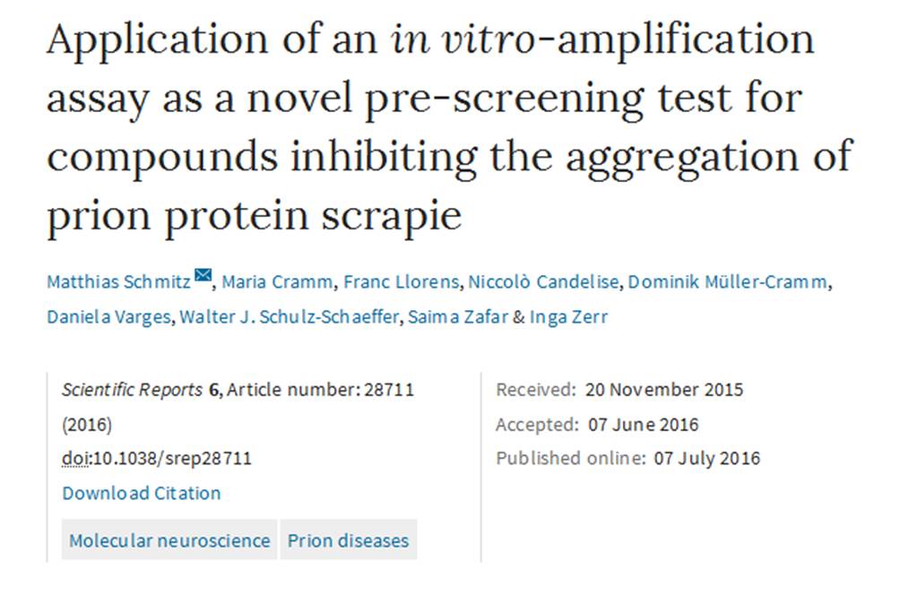 “Application of an in vitro-amplification assay as a novel pre-screening test for compounds inhibiting the aggregation of prion protein scrapie” has been published in Scientific Reports. This research was supported in part by JPND through the DEMTEST project, selected under the 2011 biomarkers call.
Ten international JPND working groups recommended for funding
The EU Joint Programme – Neurodegenerative Disease Research (JPND) has released the results of a “rapid-action” call to support working groups of leading scientists to bring forward novel approaches that will enhance the use of brain imaging for neurodegenerative disease research.
Ten working groups have been recommended for funding to address the methodological challenges facing different imaging modalities, among them MRI, PET, ultrasound, MEG and EEG, as well as multimodal approaches. The working groups cover a range of neurodegenerative diseases, including Alzheimer’s disease, Parkinson’s disease, Frontotemporal dementia and Huntington’s disease.
“Brain imaging has made enormous progress in recent years and is currently one of the most promising avenues in neurodegenerative disease research,” said Professor Thomas Gasser, Chair of the JPND Scientific Advisory Board. “If we can solve the challenges in the field, brain imaging could rapidly lead to faster and better diagnoses as well as a deeper understanding of the fundamental aspects and mechanisms of neurodegeneration.”
Although imaging techniques have brought about a dramatic improvement in the understanding of neurodegenerative diseases, there remain a number of significant challenges in the field. These include the execution of multi-centre clinical trials of an unprecedented scale, data transfer across imaging centres and the use of imaging for diagnostics and for measuring clinical outcomes.
To address these questions, on January 8, 2016, JPND launched a call for community-led working groups on harmonisation and alignment in brain imaging methods. The proposals recommended for funding are for top scientists to come together and propose, through ‘best practice’ guidelines and/or methodological frameworks, how to overcome key barriers to the use of imaging in neurodegenerative disease research.
The call attracted proposals with partners from across Europe and beyond, including Asia, Australia, North America and South America. A notable number of groups based in the United States were involved in responses to the call. Funding decisions were based upon scientific evaluation and recommendations to sponsor countries by a JPND peer review panel.
“This call perfectly embodies JPND’s mission and objectives,” said Professor Philippe Amouyel, Chair of the JPND Management Board. “The purpose of JPND is to strengthen coordination and collaboration in neurodegenerative disease research across different countries. We want to ensure that research efforts are not duplicated, to build consensus and to accelerate a path toward a cure that works. This call convenes groups of leading experts to hammer out the hard questions, including the challenges of interoperability and shared and open data, to allow researchers to more rapidly and more fully exploit imaging techniques going forward.”
Each working group is expected to run for a maximum of 9 months. The outputs of the working groups are to be produced by the end of the funding period, and will be published on the JPND website and used for further JPND actions. In addition, a common workshop will be organised to bring together and present the recommendations of each working group, encouraging the further exchange of ideas and wider dissemination to different stakeholder groups.
For more information on the working groups recommended for funding, click here.
